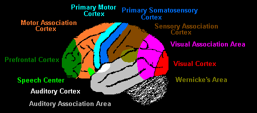
The divisions of the brain correspond with particular body functions. Even though we previously discussed the purposes of brain lobes, different parts of each brain lobe are in charge of different functions. For example, the frontal lobe controls all forms of executive action. But certain areas of the frontal lobe specialize in different forms of executive action: the primary motor cortex controls basic movement while the outer prefrontal lobe controls judgement abilities.
Brodmann Areas and Functions
There are 52 total Brodmann areas, which have been grouped into 11 areas with similar cytoarchitecture. Below are Korbinian Brodmann's actual diagrams of the brain.
 |
| Lateral Surface:Side Surface of Brain |
 |
| Medial Surface:Middle Slice of Brain |
Primary Somatosensory Cortex (Areas 1, 2, 3): Primary receptive area for touch sensations.
Primary Motor Cortex (Area 4): Communicates with spinal cord to coordinate large muscle movements.
Primary Visual Cortex (Area 17): Processes visual information, such as visual stimuli and object recognition.
Primary Auditory Cortex (Area 22): Processes auditory information, such as source of sound identification, sound recognition, and frequency recognition.
Wernicke's Area (Areas 39, 40): Produces written and spoken language, and is one of two linked speech areas in the brain (linked with Broca's Area)
Broca's Area (Areas 44, 45): Associated with more complex language production, such as inferences, complex sentences, sarcasm, etc.
The most important (for this project) Brodmann areas have been labeled. While these areas provide more detailed insight into the functions of each brain area, for the purposes of this project, I will use a simpler diagram.
Previously unidentified areas in the diagram above are explained below. Please refer to the previous set of definitions as well.
Speech Center: see Broca's Area
Pre-frontal Cortex: Coordinates concentration, judgement, and problem-solving abilities.
Motor Association Cortex: Coordinates more complex and fine movementsSensory Association Cortex: Processes multisensory information and associated them together and in context.
Visual Association Cortex: Processes visual information and associates them with memory.
Importance - Diagnosis
So what is so important about the Brodmann Areas? An understanding of the regions and their associated functions is essential to treating mental disorders. If the QEEG reports abnormalities in portions of the brain, those functions are likely to be affected as well. Excess/lack of brainwaves in those regions will either speed up or slow down associated functions of that region. For example, if excess Theta is found in the frontal lobes, a client will not be attentive or concentrated. Therefore, the client is likely to have Attention Deficit Disorder. Any noticeable behavioral problems in a client will allow a therapist to pinpoint a Brodmann area of disorder. This will result in faster detection of problems, more effective treatment specific to that problem, and an understanding of the physical problems of the client's brain.


That's pretty interesting! I also heard somewhat about Brodmann areas for my SRP and I'm glad to get a better understanding!
ReplyDelete